

 한국어
 简体中文
FACILITIES
Equipment
The equipment we use for accurate diagnosis and safe treatment.
Joy Dental Clinic Bongcheon, near Bongcheon Station, Gwanak-gu.
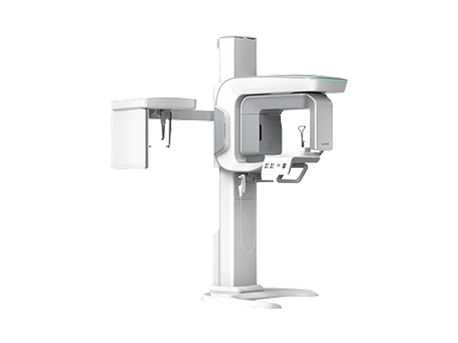
Precision diagnosis
3D CT Vatech Green Smart (PHT-35LHS)
A 3D diagnostic unit that takes panoramic, cephalometric and CBCT images in one machine
Panoramic, cephalometric and three-dimensional (CBCT) images are all taken on one unit. Structures that overlap on a single panoramic X-ray are reconstructed as cross-sections from several directions, so the height, thickness and density of the jawbone and the positions of the inferior alveolar nerve canal and maxillary sinus can be checked in advance and used to plan implants, wisdom tooth extraction and root canal treatment. The scan area (FOV) can be selected to suit the diagnostic purpose, and the model is designed for low dose.
- Panoramic, cephalometric and CBCT imaging in one unit
- Checking nerve and sinus positions and jawbone condition before implant placement
- Measuring the distance between an impacted wisdom tooth and the nerve canal
- Selecting the scan area (FOV) to match the area being diagnosed
Learn more Close
An all-in-one imaging unit
The Vatech Green Smart (PHT-35LHS) performs panoramic, cephalometric (optional), cone-beam CT and 3D model scanning in one unit. The mode needed is selected according to the purpose of the scan.
3D reconstruction with cone beam
A cone-shaped X-ray beam rotates once around the head, and a computer reconstructs the data into axial, sagittal and coronal sections and 3D images. Structures such as the teeth, periodontal ligament, alveolar bone, maxillary sinus and nerve canal can be viewed separately without overlapping.
Planning implants and root canal treatment
The height, width and density of the bone at the planned site and the distance to the inferior alveolar nerve canal and the floor of the maxillary sinus are checked to plan the position and length of the implant. It also helps to check things that are hard to see on 2D images, such as the number and curvature of roots, accessory canals and the extent of lesions at the root tip.
Scan area and radiation dose
Setting a narrow scan area (FOV) for the diagnostic purpose can reduce exposure. Whether to take a scan is decided by weighing clinical need against benefit and risk.
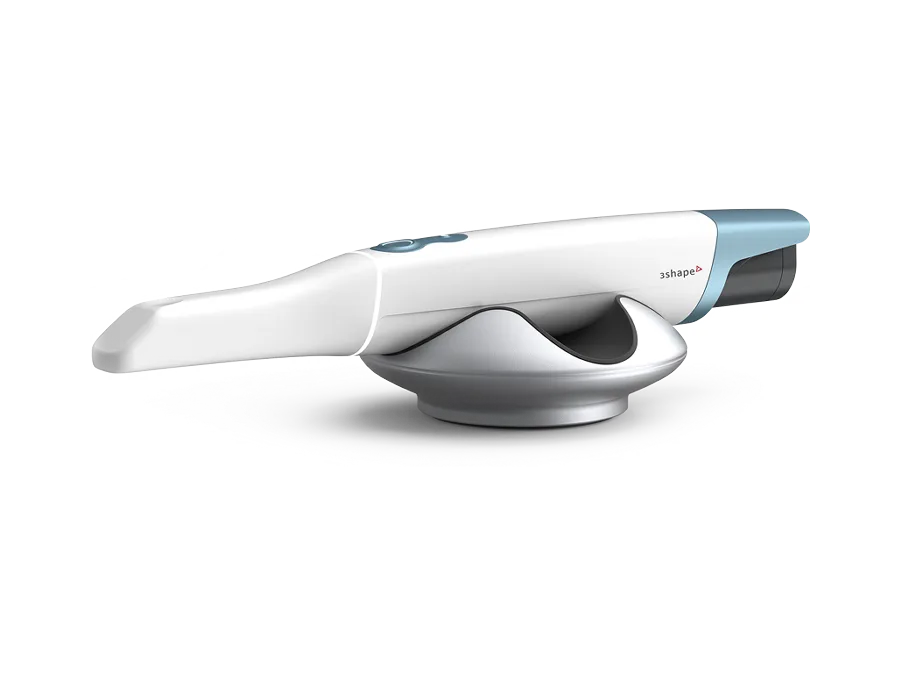
Digital prosthetics
Intraoral scanner 3Shape TRIOS 6
Scans the inside of the mouth digitally, without rubber impression material
Instead of biting into impression material and waiting for it to set, the inside of the mouth is scanned to obtain the 3D data needed to make crowns and other prostheses, orthodontic appliances and implant prostheses. The scanner is wireless, and the scanned shape can be checked on screen right away, with only the areas that need it rescanned.
- Less gagging discomfort from impression material
- Checked on screen right after scanning, with partial rescans
- Stored as data and used if a prosthesis needs to be remade
- Helps observe tooth surfaces using near-infrared and visible light
Learn more Close
Digital impressions
The inside of the mouth is scanned optically to create 3D data that replaces impression material and plaster models. The data is sent directly to prosthesis design and milling equipment, and there are no concerns about storing models or models changing shape.
Hyperspectral imaging aid
TRIOS 6 combines near-infrared, blue and white light in hyperspectral imaging to help observe the condition of tooth surfaces. A definitive diagnosis is made by considering clinical examination and X-rays together.
Wireless, calibration-free use
It is designed to be used without a separate calibration step, and it works wirelessly, which keeps the treatment workflow simple. The tips are sterilized and reused.
Explaining to patients
The scanned 3D images are used to explain your current condition and treatment plan while looking at the screen together with you.
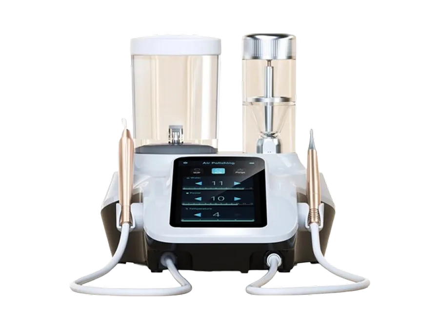
Prevention and hygiene
Air polishing (powder scaler) Woodpecker PT-B
Removes surface stains and biofilm with fine powder, water and air
Fine powder is sprayed together with water and air to remove stains and bacterial film (biofilm) left on tooth surfaces, along the gum line and around orthodontic appliances and prostheses. An ultrasonic scaler is built in, so tartar removal and stain care can be done one after the other, and low-hardness powders are used.
- Care for external stains from coffee, tobacco and the like
- Keeping areas around orthodontic appliances and implant prostheses clean
- Done together with ultrasonic scaling
- Uses low-hardness powders such as glycine and bicarbonate
Learn more Close
How air polishing works
Spraying a mix of powder, water and compressed air physically lifts deposits and biofilm off tooth surfaces. It is used for hard-to-reach areas between teeth, around orthodontic appliances and under implant prostheses.
Types of powder
Fine glycine- and erythritol-based powders are mainly used below the gum line and next to exposed roots and restorations, and sodium bicarbonate powder is mainly used to remove stains above the gum line. The powder is chosen to suit the area and the purpose.
Combined with an ultrasonic scaler
The PT-B combines a piezoelectric ultrasonic scaler and an air polisher in one unit, so stain and biofilm care can follow tartar removal in the same sitting.
Stain care vs whitening
This removes stains caused by external factors to bring teeth closer to their natural color. It is different from whitening, which lightens the shade of the tooth itself.
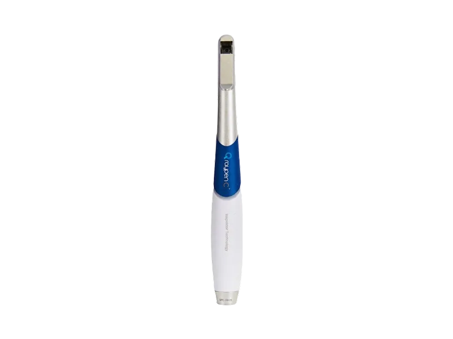
Precision diagnosis
Q-ray quantitative light-induced fluorescence Q-ray
Light of a specific wavelength makes bacterial film and early decay visible
Under 405 nm blue light, porphyrins produced by bacterial film appear red and areas that have lost minerals appear dark. Images are taken without radiation and saved as photos, and dedicated software quantifies the fluorescent area and the degree of loss so they can be compared over time.
- Imaging without radiation
- Early lesions and bacterial film checked together on photos
- Quantified and compared with fluorescence values (ΔF, ΔR)
- Observing changes before and after scaling or fluoride application
Learn more Close
How QLF works
When blue light in the visible range is shone on teeth, healthy enamel gives off green autofluorescence. Areas that have lost minerals fluoresce less and look darker, while porphyrins produced by bacterial film and mature plaque stand out as red fluorescence.
Quantifying and tracking
Dedicated software calculates values such as fluorescence loss (ΔF) and the red fluorescence ratio (ΔR), so images of the same area taken at intervals can be compared quantitatively.
What it is used for
It is used as an aid for areas suspected of early decay, checking plaque control, observing cracks, and comparing before and after scaling or professional fluoride application.
Its place in diagnosis
Fluorescence images are reference material that helps with screening and explanation. The final diagnosis is made by also considering clinical examination and X-rays.
Anesthesia comfort
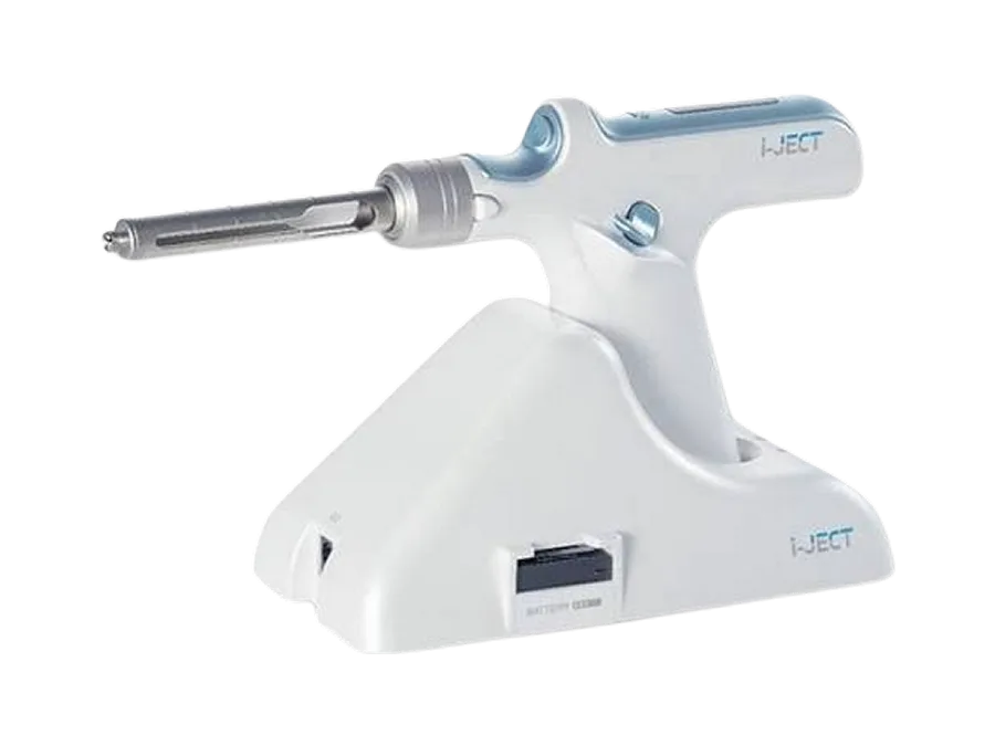
Computer-controlled anesthesia i-JECT
A machine keeps the speed and pressure of anesthetic injection steady
A built-in computer keeps the pressure steady, avoiding the pressure changes that occur with a hand-held injection, to reduce the feeling of pressure during injection. The anesthetic is warmed close to body temperature to lessen the discomfort of cold liquid going in, and an automatic aspiration function checks whether the needle is in a blood vessel. It is a Korean-made device co-developed with Seoul National University Dental Hospital.
- Injection at a steady speed and pressure
- Anesthetic warmed close to body temperature
- Automatic aspiration to check for injection into a blood vessel
- Used for patients who find injections hard and for children
Learn more Close
Computer-controlled injection
An electric drive keeps the speed and pressure of the anesthetic injection steady according to a program. The aim is to reduce the sudden pressure changes that happen when force goes into the hand during a manual injection.
Warming and automatic aspiration
The anesthetic in the cartridge is warmed close to body temperature to reduce irritation from the temperature difference. Before injecting, it automatically aspirates to check whether the needle tip has entered a blood vessel.
Measured dosing
Injection is controlled to the set amount, which helps keep the amount used consistent.
Individual differences
Manufacturer data report less pain and discomfort during injection than with a manual method, but how much difference you actually feel varies by site and by person.
Aesthetic
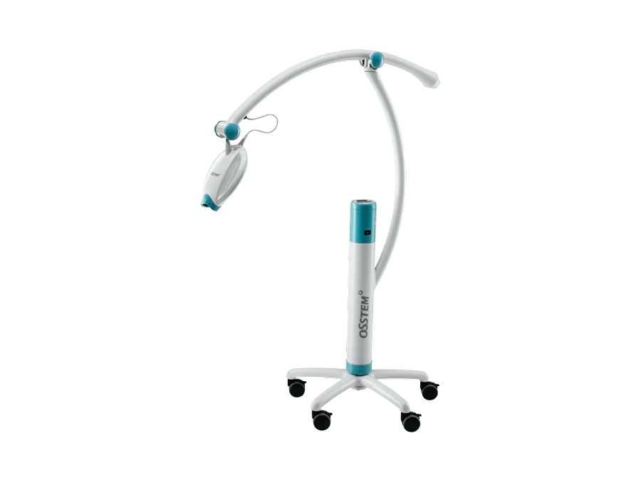
In-office whitening light Osstem BeauTis Light
A light-curing unit for professional whitening done in the clinic
After whitening gel is applied to the teeth, the whitening process is carried out with the unit’s blue LED light (in the visible range, not ultraviolet). A protective treatment for the gums is done at the same time, and the degree of color change and how long it lasts vary from person to person. It is sometimes combined with at-home whitening.
- Professional whitening done in the clinic
- Uses a visible-light LED, not ultraviolet
- Gum protection done at the same time
- Results and how long they last vary by person
Learn more Close
What the light does
It shines blue light of about 420–520 nm onto the whitening gel to assist the reaction. It does not use ultraviolet light, and protection is placed between the light and the gums.
How it is done
Hydrogen peroxide or carbamide peroxide whitening gel is applied to the tooth surfaces and light is shone for a set time, repeated over several rounds. If needed, at-home tray whitening is added to help maintain the result.
Differences in response
How teeth respond depends on the cause of discoloration (internal or external), the starting tooth color and the condition of the enamel. Prostheses and fillings do not whiten.
Symptoms during treatment
Temporary sensitivity or gum irritation can occur during whitening and usually settles over time. This varies by person and is explained at the consultation.
Precision treatment
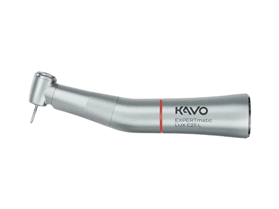
Electric handpiece KaVo EXPERTmatic LUX E25 L
An electrically driven handpiece that keeps rotation speed and torque steady
Unlike air-driven handpieces, it is driven by an electric motor, so it keeps the set rotation speed and turning force (torque) steady even under load. A 1:5 speed-increasing contra-angle with glass-fiber LED lighting lets the dentist see the area being prepared clearly, and it is used to control the rotation of root canal files precisely and to prepare teeth evenly for prostheses. Ceramic bearings make noise and vibration relatively low.
- Rotation speed and torque held steady under load
- 1:5 speed-increasing contra-angle with LED lighting to see the area being prepared
- Precise control of root canal file rotation
- Relatively low noise and vibration with ceramic bearings
Learn more Close
Steady speed and torque from an electric drive
An electric motor delivers steady torque across a wide range of speeds. Speed does not drop suddenly when pressure is applied during preparation, which helps shape the tooth evenly to the planned form.
1:5 speed-increasing contra-angle
The EXPERTmatic LUX E25 L increases the motor speed five times. With a small head and glass-fiber LED lighting (about 25,000 lux), it helps with visibility in hard-to-reach areas such as the back teeth.
Root canal shaping
In root canal shaping with nickel-titanium files, rotation speed, torque and rotation angle are set finely to manage the load on the file.
Noise, vibration and care
Ceramic ball bearings keep the operating sound low at about 57 decibels, with little vibration. It can be sterilized at high temperature, which makes hygiene management easier.
Treatment environment
Shadowless operating light LED shadowless light
A treatment light that reduces shadows to light the treatment area evenly
Multiple LED light sources with reflector and lens design reduce shadows cast by hands or instruments. A color temperature close to natural light and a high color rendering index (CRI) are used so colors can be told apart accurately, and brightness and color temperature can be adjusted. There is little heat from ultraviolet or infrared light.
- Fewer shadows from instruments and hands
- Color temperature close to natural light helps tell colors apart
- Adjustable brightness and color temperature
- LED light source with little heat
Learn more Close
How a shadowless light works
Several light sources and reflective surfaces facing different directions are combined so that when one source is blocked, light from the others fills in the shadow. The whole treatment area is lit evenly.
Color temperature and color rendering
A color temperature of around 5,000 K and a high color rendering index (CRI) let the colors of gums, teeth and restorations be checked close to how they look in natural light. This helps when choosing the shade of a prosthesis.
Adjustment and heat
Brightness and color temperature are adjusted to the treatment being done. Because it is LED, it gives off little ultraviolet, infrared or heat, so it is easy to use for long periods.
※ Equipment names and models are those actually in use and may change with the time of introduction. Whether a device is used, and the specific treatment method, are decided after consultation according to the patient’s condition.
Book a detailed diagnostic consultation
Equipment is a tool that supports accurate diagnosis. A treatment plan suited to your symptoms and condition is explained at the consultation.
Book a consultation